
The brain contains an extraordinary variety of cells organized into highly specialized anatomical regions. A molecular change occurring in one neuronal population may have very different implications from the same change occurring elsewhere. This makes location especially important when researchers investigate neurological disease.
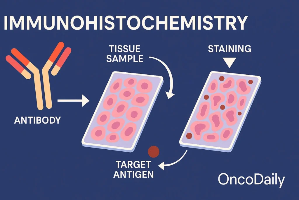
Immunohistochemistry (IHC) allows scientists to detect specific proteins while preserving the structure of brain and nervous-system tissue. Researchers can use immunohistochemistry services to investigate protein localization, neuronal populations, glial responses, inflammation, and pathological changes while maintaining the spatial context needed to interpret neurological biology.
Why Is Spatial Biology Important in Neuroscience?
The nervous system is highly organized.
Different regions of the brain perform different functions, and individual regions contain multiple cell populations with specialized roles.
A molecular assay performed on homogenized tissue can reveal that a protein is present or altered, but it may not identify which cells express it.
For neuroscience research, this distinction can be critical.
A protein detected in neurons may have a different biological meaning from the same protein detected in astrocytes, microglia, or vascular cells.
IHC helps connect molecular expression with anatomy.
Identifying Different Neural Cell Populations
Brain tissue contains more than neurons.
Researchers may need to distinguish among several major cell types, including:
- Neurons
- Astrocytes
- Microglia
- Oligodendrocytes
- Endothelial cells
Each population contributes differently to nervous-system function and disease.
By selecting appropriate markers, researchers can identify these cells within intact tissue and examine how their distribution or characteristics change under different experimental conditions.
Studying Neurons Within Their Anatomical Context
Neurons form complex networks extending across different regions of the nervous system.
Researchers may use IHC to examine proteins associated with neuronal identity, signaling, structure, or disease.
Preserving tissue architecture makes it possible to investigate whether molecular changes occur broadly throughout the brain or are concentrated in particular anatomical areas.
This can be useful when studying disorders in which specific neuronal populations are disproportionately affected.
Microglia and Neuroinflammation
Microglia are resident immune cells of the central nervous system.
They respond to changes in their local environment and are widely investigated in research involving neuroinflammation, injury, infection, and neurodegenerative disease.
Researchers can use tissue markers to examine microglial distribution and changes associated with experimental conditions.
The location of these cells can provide useful context.
For example, researchers may investigate whether microglia accumulate around areas of tissue damage or near pathological protein deposits.
Understanding the Role of Astrocytes
Astrocytes perform numerous functions within the nervous system, including supporting neurons and helping maintain the local cellular environment.
They can also undergo substantial changes following injury or during disease.
IHC allows researchers to visualize astrocyte-associated markers while retaining their relationship to neurons, blood vessels, and other tissue structures.
This spatial information can help researchers investigate how astrocyte responses differ across brain regions or experimental groups.
Investigating Neurodegenerative Disease
Neurodegenerative diseases involve progressive changes in particular neuronal systems and are often associated with abnormal protein accumulation, inflammation, and cellular dysfunction.
Tissue-based analysis can help researchers investigate these processes simultaneously.
Depending on the research question, IHC may be used to examine:
- Pathological protein accumulation
- Neuronal loss
- Microglial responses
- Astrocyte activation
- Synaptic proteins
- Cellular stress markers
Examining these features within the same anatomical context can help researchers understand relationships between molecular pathology and tissue changes.
Protein Aggregation and Localization
Several neurological diseases involve proteins that misfold or accumulate abnormally.
Simply measuring total protein abundance may not reveal whether a protein has changed its cellular localization or formed abnormal deposits.
IHC can help visualize where these proteins occur within tissue.
Researchers may investigate whether staining appears:
- Inside neurons
- Outside cells
- Around blood vessels
- In particular brain regions
- Near inflammatory cells
These patterns can provide additional information about disease-associated processes.
IHC in Brain Tumor Research
Neurological tissue research also includes tumors of the central nervous system.
Brain tumors can contain heterogeneous populations of malignant cells along with immune, vascular, and stromal components.
Researchers may use IHC to characterize proteins associated with:
- Tumor identity
- Cell proliferation
- Signaling pathways
- Immune infiltration
- Vascular structures
- Experimental therapeutic targets
Spatial analysis can help distinguish tumor cells from surrounding brain tissue and identify regional differences within the tumor itself.
Studying the Blood-Brain Barrier
The blood-brain barrier regulates movement between the bloodstream and the central nervous system.
Its function depends on interactions among endothelial cells, supporting cells, and specialized molecular structures.
Researchers can use tissue staining to investigate proteins associated with barrier integrity and vascular organization.
This can be relevant in studies involving neurological disease, inflammation, injury, and experimental drug delivery.
Why Tissue Preparation Matters
Brain tissue presents particular challenges for histological analysis.
Fixation needs to preserve morphology while maintaining sufficient antigen accessibility for antibody detection.
Several factors can affect staining quality:
- Fixation conditions
- Section thickness
- Antigen retrieval
- Antibody concentration
- Detection method
- Tissue handling
Consistency becomes particularly important when comparing samples from different experimental groups.
Poor tissue preparation can introduce variability that is difficult to distinguish from genuine biological differences.
Antibody Specificity Is Critical in Brain Research
The nervous system contains thousands of proteins, including closely related receptors, channels, enzymes, and structural molecules.
An antibody that cross-reacts with unrelated proteins can produce misleading staining patterns.
Researchers should therefore consider evidence supporting antibody specificity for the intended tissue and application.
Useful validation approaches may include:
- Positive controls
- Negative controls
- Knockout or knockdown tissue
- Expected anatomical distribution
- Comparison with independent methods
A visually convincing image is not sufficient evidence of target specificity on its own.
Quantifying Neurological Tissue Changes
Modern IHC studies can move beyond qualitative descriptions.
Digital imaging enables researchers to quantify tissue characteristics such as:
- Number of positive cells
- Staining intensity
- Area occupied by a marker
- Distribution of pathological deposits
- Distance between cellular populations
Standardized image-analysis protocols can improve comparisons across treatment groups or disease models.
However, automated analysis should be checked carefully because complex neural structures can create segmentation challenges.
Multiplex Analysis in Neuroscience
Studying one marker at a time can provide limited information about interactions among different neural cell types.
Multiplex staining allows researchers to visualize several targets within the same tissue section.
For example, researchers could examine a neuronal marker together with markers for microglia, astrocytes, and a disease-associated protein.
This makes it possible to investigate whether inflammatory cells are located near affected neurons or pathological deposits.
Spatial relationships of this kind can generate hypotheses that would be difficult to derive from separate tissue sections.
Connecting Histology With Omics Data
Neuroscience increasingly uses genomics, transcriptomics, proteomics, and single-cell technologies.
These approaches can identify molecular patterns associated with neurological conditions, but researchers often need tissue-level methods to determine where those signals occur.
A study might use transcriptomic analysis to identify a protein associated with disease and then use immunohistochemistry services to investigate whether that protein is localized to neurons, glial cells, vascular structures, or particular anatomical regions.
This combination connects large-scale molecular discovery with visible tissue biology.
IHC in Preclinical Neurological Research
Animal models remain important for investigating neurological mechanisms and potential therapies.
Researchers can compare brain tissue from treated and untreated groups to determine whether an intervention affects selected pathological or cellular markers.
Tissue analysis may complement behavioral measurements, imaging, molecular assays, and other endpoints.
For example, an experimental treatment might improve a behavioral outcome while also changing neuroinflammatory markers within a particular brain region.
Combining these observations provides more biological context than either measurement alone.
What Should Researchers Plan Before an IHC Study?
A successful neuroscience IHC experiment begins with a clearly defined question.
Researchers should consider:
- Which cell population is relevant?
- Which brain region should be examined?
- What protein or biological process is being measured?
- Is the antibody validated for the species and tissue?
- Which controls are required?
- How will staining be quantified?
- Are multiple markers needed?
These decisions should be made before staining begins because they influence tissue collection, sectioning, antibody selection, imaging, and analysis.
Looking Ahead
Modern neuroscience increasingly combines molecular information with spatial biology.
Sequencing can identify cellular states, proteomics can reveal changes in protein abundance, and computational methods can uncover patterns across large datasets. Yet researchers still need to know where these changes occur within the highly organized nervous system.
Immunohistochemistry provides that connection.
As multiplex imaging, digital pathology, spatial molecular technologies, and computational analysis continue to develop, researchers will be able to investigate neural tissue with increasing detail.
The result is a more integrated view of neurological disease, one that connects molecular changes with specific cells, anatomical regions, and pathological processes.
